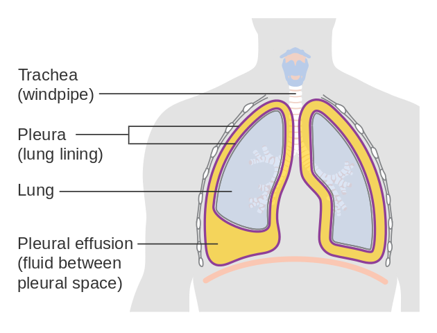
Loculated Pleural Effusion : Pleural Effusion : 1 pleural effusion is defined as abnormal fluid collection in the pleural space.. Most pleural effusions, whether free flowing or loculated, are hypoechoic with a sharp echogenic line that delineates the visceral pleura and lung. Complex septated, complex nonseptated, or homogeneously echogenic effusions are always exudates (fig. Loculated pleural effusion the pleura is a thin membrane between the lungs and chest wall that lubricates these surfaces and allows movement of the lungs while breathing. Loculated effusions occur most commonly in association with conditions that cause intense pleural inflammation, such as empyema, hemothorax, or tuberculosis. Causes of an exudative effusion are malignancy, infection, or inflammatory disorders such as rheumatoid arthritis.
A pleural effusion is due to the manifestations of another illness. A pleural effusion representsthe disruption of the normal mechanisms of formationand drainage of fluid from the pleural space. Nine of the 19 malignant effusions showed loculation (47%). Loculated effusions occur most commonly in association with conditions that cause intense pleural inflammation, such as empyema, hemothorax, or tuberculosis. Pleural effusion that is confined to one or more fixed pockets in the pleural space.
Xmlinkhub from e-kcj.org Tube thoracostomy has variable success in the treatment of complex pleural effusions, with Empyema and large or loculated effusions need to be fo … at least 40% of all patients with pneumonia will have an associated pleural effusion, although a minority will require an intervention for a complicated parapneumonic effusion or empyema. Nonmalignant pleural effusions (nmpes) have a wide variety of etiologies ( table 1 and table 2 and table 3) and cause significant morbidity and mortality 2,3 . Initial testing … lupus pleuritis and other causes of pleural effusions in lupus patients. Pleural effusion that is confined to one or more fixed pockets in the pleural space. Treatment may fail if the catheter is not placed optimally within the loculation or if the fluid is hemorrhagic or fibrinous. Of loculated pleural effusions* jeffreys. We studied the value of transca …
Most malignant effusions can be controlled by thoracentesis and/or closed thoracostomy tube drainage and sclerosis of the pleural cavity.
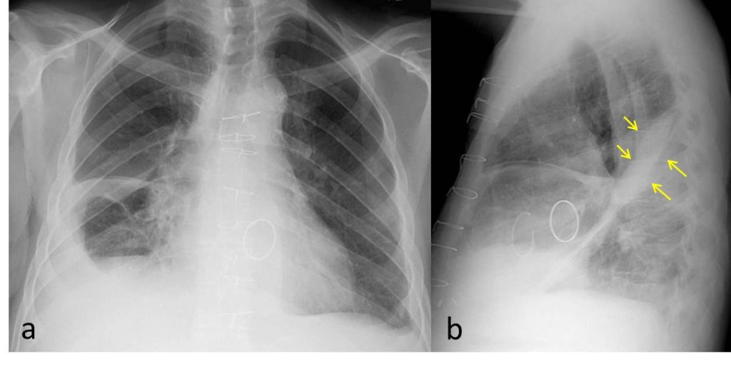
Loculated malignant effusions however, are inherently resistant to the usual approaches because of nonexpanding underlying lung. The largest pocket of fluid is present posteriorly at the right lung base, with associated atelectasis and minor consolidation. Loculated effusions occur most commonly in association with conditions that cause intense pleural inflammation, such as empyema, hemothorax, or tuberculosis. Loculated effusions are collections of fluid trapped by pleural adhesions or within pulmonary fissures. Most pleural effusions, whether free flowing or loculated, are hypoechoic with a sharp echogenic line that delineates the visceral pleura and lung. A pleural effusion representsthe disruption of the normal mechanisms of formationand drainage of fluid from the pleural space. An anechoic effusion can be a transudate or exudate (fig. Treatment may fail if the catheter is not placed optimally within the loculation or if the fluid is hemorrhagic or fibrinous. We studied the value of transca … Surgical treatment of pleural effusion may include chest. Causes of an exudative effusion are malignancy, infection, or inflammatory disorders such as rheumatoid arthritis. If the fluid cannot be drained, the lungs aren't able to expand and oxygenate the blood sufficiently. Pleural effusion predominantly presents with breathlessness, but cough and pleuritic chest pain can be a feature.
Loculated malignant effusions however, are inherently resistant to the usual approaches because of nonexpanding underlying lung. The lack of specificity is mainly due to the limitations of the imaging modality. The reasons for effusion are many, and the specific diagnosis is often based upon tap or drainage of the fluid. A pleural effusion representsthe disruption of the normal mechanisms of formationand drainage of fluid from the pleural space. A pleural effusion is due to the manifestations of another illness.
Imaging Findings Of Free Flowing Effusion Online Medical Library from d3uigcfkiiww0g.cloudfront.net Most effusions start like this and can be easily missed. Tube thoracostomy has variable success in the treatment of complex pleural effusions, with Nonmalignant pleural effusions (nmpes) have a wide variety of etiologies ( table 1 and table 2 and table 3) and cause significant morbidity and mortality 2,3 . In vitro efficacy of varidase versus streptokinase or urokinase for liquefying thick purulent exudative material from loculated empyema. Surgical treatment of pleural effusion may include chest. Loculated pleural effusion the pleura is a thin membrane between the lungs and chest wall that lubricates these surfaces and allows movement of the lungs while breathing. Empyema and large or loculated effusions need to be fo … at least 40% of all patients with pneumonia will have an associated pleural effusion, although a minority will require an intervention for a complicated parapneumonic effusion or empyema. Surgical thoracostomy tube placement and radiologically guided catheter drainage are standard therapy for loculated pleural fluid collections.
In vitro efficacy of varidase versus streptokinase or urokinase for liquefying thick purulent exudative material from loculated empyema.
Lung scarring and a permanent decrease in lung function are associated with chronic pleural effusion. This type of effusion is empyema unless proven otherwise. A pleural effusion representsthe disruption of the normal mechanisms of formationand drainage of fluid from the pleural space. Pleural effusion predominantly presents with breathlessness, but cough and pleuritic chest pain can be a feature. A pleural effusion occurs when fluid fills this gap and separates the lungs from the chest wall. Icu patients cannot sit up and the effusion layers posteriorly. In vitro efficacy of varidase versus streptokinase or urokinase for liquefying thick purulent exudative material from loculated empyema. The largest pocket of fluid is present posteriorly at the right lung base, with associated atelectasis and minor consolidation. Pleural effusion that is confined to one or more fixed pockets in the pleural space. Surgical thoracostomy tube placement and radiologically guided catheter drainage are standard therapy for loculated pleural fluid collections. Loculated pleural effusion the pleura is a thin membrane between the lungs and chest wall that lubricates these surfaces and allows movement of the lungs while breathing. The pleural space is normally filled with ~5 to 10 ml of serous fluid, which is secreted mainly from the parietal pleura at a rate of 0.01 ml/kg/h and absorbed through the lymphatics. If it is clear that there are multiple loculations then it is wise to avoid delay and proceed directly to this procedure.
Surgical thoracostomy tube placement and radiologically guided catheter drainage are standard therapy for loculated pleural fluid collections. Tube thoracostomy has variable success in the treatment of complex pleural effusions, with The lack of specificity is mainly due to the limitations of the imaging modality. 1 pleural effusion is defined as abnormal fluid collection in the pleural space. Pleural effusion that is confined to one or more fixed pockets in the pleural space.
Epos Trade from epos.myesr.org A pleural effusion occurs when fluid fills this gap and separates the lungs from the chest wall. 681 views reviewed >2 years ago Icu patients cannot sit up and the effusion layers posteriorly. (vats) with lysis of adhesions is also a viable option for loculated effusions. Pleural fluid is seen extending to the right oblique fissure. Nine of the 19 malignant effusions showed loculation (47%). Most pleural effusions, whether free flowing or loculated, are hypoechoic with a sharp echogenic line that delineates the visceral pleura and lung. Left pleural effusion with high density material at the posterior costophrenic angle.
Loculated pleural effusion the pleura is a thin membrane between the lungs and chest wall that lubricates these surfaces and allows movement of the lungs while breathing.
Nine of the 19 malignant effusions showed loculation (47%). Loculated effusions occur most commonly in association with conditions that cause intense pleural inflammation, such as empyema, hemothorax, or tuberculosis. Surgical treatment of pleural effusion may include chest. The lack of specificity is mainly due to the limitations of the imaging modality. Of the 22 transudates, eight showed a loculated pleural effusion (36%) compared with 45 of 78 exudates (58%). This type of effusion is empyema unless proven otherwise. The pleura are thin membranes that line the lungs and the inside of the chest cavity and act to lubricate and facilitate breathing. Pleural effusions are very common, and physicians of allspecialties encounter them. Initial testing … lupus pleuritis and other causes of pleural effusions in lupus patients. Pleural effusions describe fluid between the two layer of tissue (pleura) that cover the lung and the lining of the chest wall. A pleural effusion is due to the manifestations of another illness. A pleural effusion occurs when fluid fills this gap and separates the lungs from the chest wall. Tube thoracostomy has variable success in the treatment of complex pleural effusions, with


0 Komentar